


以下内容原文发布于AACR官方博客《Cancer Research Catalyst》, 中文内容仅做参考,请点击文末“阅读原文”,阅览原文内容。
临床层面的突破性进展始于实验室工作台上的基础研究,而实验室研究又依赖于技术进步,以此支持科学家重新认识问题并为患者找到新的解决方案。如果说全方位的癌症研究是一台机器,那么上述三个层面共同发挥作用才能让它高效运转。
2024年美国癌症研究协会(AACR)年会于4月5日至10日在圣迭戈举行,大会日程委员会主席希望将“技术—实验室研究—临床”这一范式作为本届年会的基础。
斯坦福大学医学、遗传学和生物医学数据科学RZ Cao教授、人工智能与癌症基因组学系主任、乳腺癌转化研究主任Christina
Curtis博士在此次年会的开幕全体会议上致辞称:“(我们通过)共同努力,将基础科学、转化科学和临床科学领域相关成果的报告与支持和扩大这些成果的工具相关的报告搭配展示,从而让技术融入科学计划。”
马萨诸塞州综合癌症中心临床研究主任、哈佛大学医学院医学系教授Keith
T. Flaherty博士(MD、AACR会士)与Christina
Curtis博士共同主持了本届年会的日程委员会和开幕全体会议。二人共同工作了一整年,以指导会议的重点和范围。在欢迎与会者参加开幕全体会议时,他们强调了用跨学科方法解决复杂问题的重要性,以及他们如何从这一角度出发设计会议日程。
Flaherty博士称:“我们希望能超越标准化的多学科研讨会,取而代之的是展示多学科方法如何相互启发的专题研讨会——例如对临床研究、计算方法和新型诊断方法的综合展示,或是重点介绍基础科学进展如何借助计算科学应用于临床。”
此次开幕全体会议正是这样一个将技术、实验室发现和临床进展相互融合的生动实例。会议的主题是“激励科学、推动进步、革新医疗”,四位报告人以专家角度阐述了对人体最微小单位的研究如何为临床医疗带来巨大进步。
了解更多内容,请阅读以下原文。
Opening Plenary Showcases Molecular Advances Leading to Big Revolutions in Cancer Research
Groundbreaking clinical developments begin with fundamental research at the lab bench. Bench research depends on technological advances to allow scientists to reconceptualize problems and find new solutions for patients. The full spectrum of cancer research encompasses all of these facets working together as a well-oiled machine.
It’s a paradigm that the program chairs of the American Association for Cancer Research (AACR) Annual Meeting 2024 , held April 5-10 in San Diego, wanted at the foundation of the meeting.
“[We made] a concerted effort to weave technology into the scientific program by pairing presentations on discoveries in basic science, translation, and clinical science with presentations on the tools that support and amplify those discoveries,” said Christina Curtis, PhD , the RZ Cao Professor of Medicine, Genetics, and Biomedical Data Science; director of Artificial Intelligence and Cancer Genomics; and director of Breast Cancer Translational Research at Stanford University, during the Opening Plenary session of the meeting.
Curtis chaired the Program Committee and the Opening Plenary session with Keith T. Flaherty, MD, FAACR , director of Clinical Research at Mass General Cancer Center and a professor of medicine at Harvard Medical School. Together, the two have worked for a full year to direct the focus and scope of the meeting. As they welcomed attendees to the Opening Plenary session, they emphasized the importance of interdisciplinary approaches to complex problems, and how they designed the meeting program with that angle in mind.
“We hoped to move beyond standard multidisciplinary sessions and instead create sessions that demonstrate how these different approaches inform one another—sessions that, for example, showcase clinical investigations, computational approaches, and a novel diagnostic, or sessions that highlight basic science advances whose clinical application is being informed by computational insights,” Flaherty said.
The Opening Plenary acted as a poignant example of this intermingling of technology, laboratory discoveries, and clinical advances. In an expert reflection of the meeting theme—inspiring science, fueling progress, revolutionizing care—the four speakers shed light on how research on the tiniest elements of the human body can indeed inform gargantuan advances in clinical care.
MAPPING THE ROUTE FROM MOLECULES TO DRUGS
Just as geographical atlases depict roads, businesses, and other points of interest and help represent the relationships between them, cell atlases provide a high-resolution map of how individual cells with distinct functions are distributed throughout a tumor, tissue, or organism.
Aviv Regev, PhD, FAACR , head and executive vice president of Research and Early Development at Genentech, is head of the Human Cell Atlas project, which aims to map all of the cells in the body to better understand human development and disease.
So far, the Human Cell Atlas has mapped several million cancer cells across a variety of tumor types, Regev said. Current challenges involve analyzing this vast library of data in ways that can benefit patients.
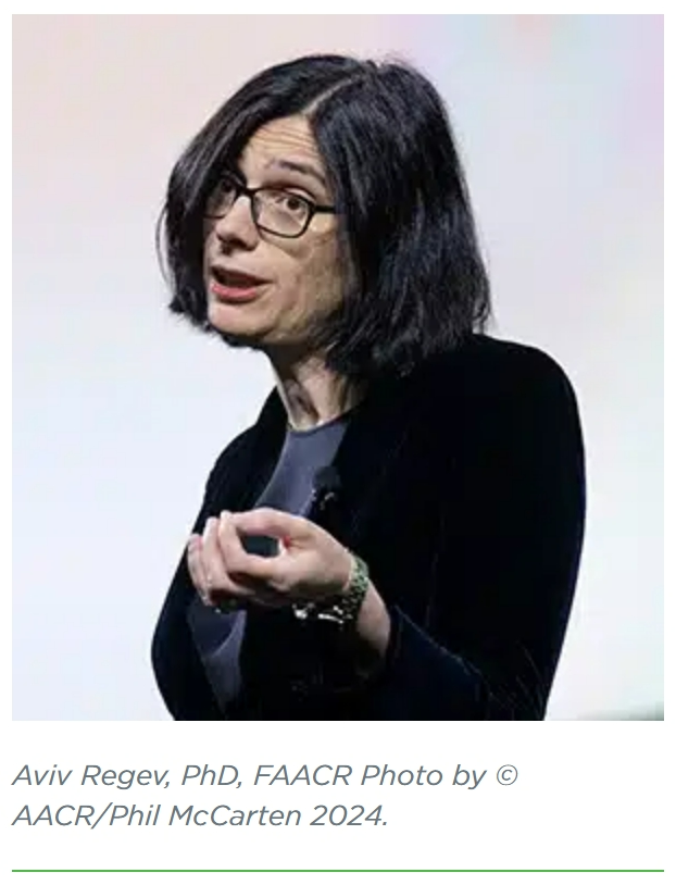
Regev likened the data generated by older genomics methods to a fruit smoothie—information from multiple cell types blended together making individual origins indistinguishable. Tech advances in single-cell genomics have allowed researchers to look at tissue in a more granular manner, similar to examining each piece of fruit in great detail.
Insights from skin cells have helped researchers study melanoma progression and develop possible treatment strategies—especially for tumors that respond poorly to immunotherapy. A combination of spatial analyses and single-cell RNA sequencing revealed that malignant cells comprising immune “cold” niches have a distinct gene expression signature that can predict immune cell exclusion. Regev and colleagues found that while tumors with more “cold” niches did not respond as well to immunotherapy as those with more “hot” niches, they were more likely to respond to inhibitors of the cyclin dependent kinases 4 and 6 (CDK4/6), suggesting a potential therapeutic vulnerability.
Regev touched on some newer technologies that can facilitate the comprehensive mapping of tumors, including ways that artificial intelligence (AI) and machine learning can help recognize patterns in the data. She explained the “lab in a loop” system of discovery, in which vulnerabilities uncovered during high-throughput screening can inform new drugs to be tested in the clinic, which can, in turn, provide real-world data that may raise questions for new screens.
AI can assist with many steps of this process, Regev said. She showed how machine learning could help streamline perturbation screens , for instance; instead of disrupting a single gene per cell, researchers may now be able to disrupt multiple genes in tandem, allowing the AI to look for patterns that can discern the effects of each perturbation. When such screens lead to potential drug targets, new machine learning algorithms can also help predict which putative compounds have the highest chance of turning into successful drugs.
“Drug discovery and development, just like biology, only work when all components are tied together,” Regev said. “We rely on everyone and everything being connected together—the lab, the clinic, the data, and the algorithms—but when they are connected together, they have the potential to go after the big problems.”
ROBOT READERS: AI IN HISTOPATHOLOGY
Jakob Nikolas Kather, MD , a professor of clinical artificial intelligence at Technical University Dresden in Germany, was equally enthusiastic about the ways researchers are using new AI technologies to potentially enhance clinical care.
“For artificial intelligence, we need a lot of data, and one type of data that we really like is routinely available data. When we treat our cancer patients in clinical routine, a lot of data is generated as a byproduct,” Kather said.

While physicians can routinely assess genomic characteristics and expression of various proteins in patient tumors—markers that may help predict prognosis or direct treatment options—these tests require resources and infrastructure that aren’t available everywhere. Conversely, histopathology slides stained with hematoxylin and eosin (H&E) are cheaper and readily available. Can researchers infer complex biomarker information from a simple stained slide?
Kather and colleagues have shown that they can feed histopathology slides into a deep learning model and train the model to recognize which tumors exhibit high microsatellite instability, a marker of eligibility for certain therapies. They have also trained AI models to predict the prognosis of patients with colorectal cancer and determine —without staining for PD-L1—if the tumor might respond to PD-1 or PD-L1 inhibitors.
“The hope is that we can use these biomarkers on very inexpensive, routinely available H&E data,” Kather said. “Of course, to bring this to the clinic, we have to take many steps … but the technology is here, and now it’s basically our job to bring it … to our patients.”
Kather touched on new types of machine learning offering potential fresh approaches to process data. These include multimodal models that incorporate basic H&E data with immunohistochemistry or genomic data, as well as foundation models that use a two-step approach to let the algorithm recognize broad patterns in data before telling it what it needs to differentiate. He also discussed how large language models—such as ChatGPT—are beginning to use insights from language processing to analyze images and electronic health records.
“We’re living in very exciting times, and basically, our job is to take these new technologies and to translate them into new scientific insights and clinical usefulness for cancer patients,” Kather said.
GOING GLOBAL: NOVEL INSIGHTS INTO TARGET AND DRUG DISCOVERY
What happens when the insights gleaned from large data sets and AI highlight potential targets we don’t yet know how to drug?
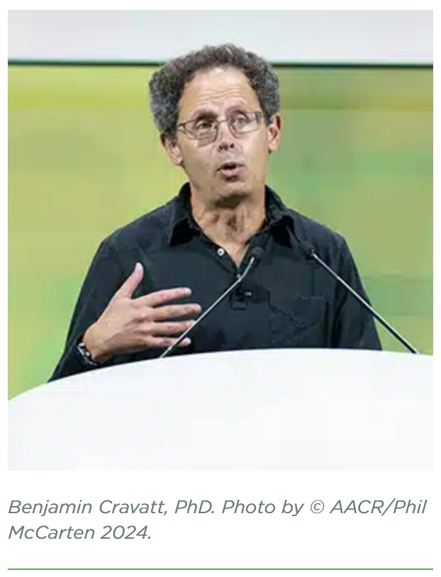
“The genetic methods point us to proteins and pathways that remain poorly characterized biochemically, that lack chemical tools and might even be considered historically undruggable,” said Benjamin Cravatt, PhD , a professor and the Norton B. Gilula Chair in Biology and Chemistry at the Scripps Research Institute. Cravatt emphasized the importance of finding new drug targets by mapping so-called “ligandability” across the proteome.
Cravatt and colleagues designed a method called activity-based protein profiling to probe for all the places in the proteome that a potential drug structure might bind. Fluorescent probes are designed to mimic ligands that can target binding pockets of interest throughout the proteome. Protein lysates are incubated first with the probes, then with a putative drug compound, to assess which drugs might outcompete the probes in proteins of interest.
This method was successfully used to identify compounds that can target serine hydrolases, a metabolic enzyme that is upregulated in a variety of cancer types. Cravatt showed that one such compound could block palmitoylation of NRAS in acute myeloid leukemia cells.
In addition to targeting specific enzyme families, Cravatt explored how to perform larger-scale screening to identify potentially druggable sites across the proteome, including in proteins previously thought to be undruggable. For this, he and his colleagues assembled a library of compounds known to interact with cysteines, a common interaction site for drugs that bind covalently to proteins. The researchers incubated the library with proteins then added a cysteine-binding probe. Cysteines that did not take up the probe were assumed to have interacted with a drug compound.
Using this method, Cravatt and colleagues identified a variety of putative targets. “We got our glimpses of the power of covalent chemistry to expand ligandable—if not druggable—space,” Cravatt said. “The proteins that harbored these cysteines, for the vast majority of them, this was their first evidence of interacting with small molecules.”
They identified a cysteine binding site on the transcription factor FOXA1, a pioneer factor that helps other, cancer-driving transcription factors bind to chromatin and activate oncogenic gene programs. The researchers have designed a ligand for this difficult-to-drug protein and shown that the ligand disrupts the ability of FOXA1 to bind to its intended target sites. While they have not yet proven that their compound acts as an inhibitor, Cravatt expressed hope that they could use this data to design FOXA1 antagonists.
MOWING THE GLYCAN LAWN TO FIGHT CANCER
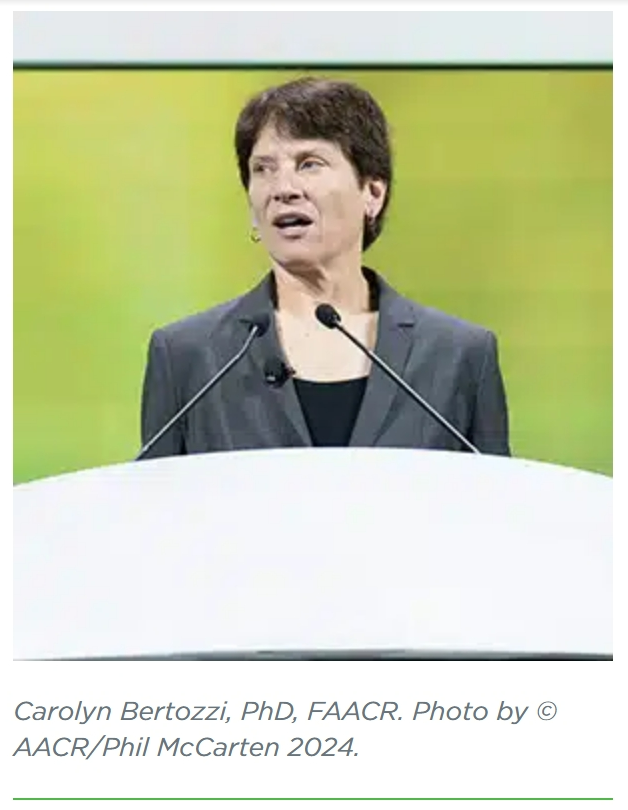
Proteins aren’t the only druggable targets in cancer cells. Carolyn Bertozzi, PhD, FAACR , the Baker Family Director of Stanford ChEM-H, the Anne T. and Robert M. Bass Professor in the School of Humanities and Sciences, and professor, by courtesy, of Chemical and Systems Biology and of Radiology at Stanford University, has turned her sights to a relatively unconventional drug target: sugars.
The cell surface is dotted with glycans—molecular scaffolds consisting of a protein, lipid, or RNA backbone complexed with various sugar tags. Sialoglycans, which contain the sugar moiety sialic acid, are commonly upregulated in tumors.
“The well-manicured garden of a healthy cell’s sialoglycans turns into a tropical forest, where they are denser and presented on different kinds of scaffolds,” Bertozzi explained.
Research has shown that sialic acid can activate an immune checkpoint-like signaling cascade in immune cells that dampens antitumor immune responses. This introduced sialoglycans as a promising drug target, but blocking their signaling proved to be more complex than blocking other immune checkpoints. The sialic acid receptors, called siglecs, form a broad family of proteins that cannot be targeted by a single antibody.
Bertozzi and colleagues pivoted to targeting the sialoglycans instead.
“The idea was to develop a therapeutic molecule that we analogized to a lawnmower , and this lawnmower would park on the surface of the targeted cancer cells and mow the lawn—and literally strip the sialic acids off of that cell, thereby depriving it of the ability to engage any member of the siglec receptor family,” Bertozzi said.
Bertozzi and colleagues designed a cancer-targeted sialidase—an enzyme that cleaves sialic acid residues—using her Nobel Prize-winning bio-orthogonal chemistry approach. The researchers ligated the sialidases to the antibody trastuzumab (Herceptin)—which targets the growth factor receptor HER2, overexpressed in a variety of human cancers—at carefully curated locations that would not disrupt the activity of trastuzumab. In mouse models, these “T-sia” molecules inhibited the growth of HER2-positive breast cancer cell xenografts.
“Attaching this sialidase to trastuzumab turns these tumors from cold to hot and provokes an immune reaction that actually is durable,” Bertozzi said.
Bertozzi and colleagues are currently streamlining their T-sia prototype to translate it to the clinic, and they aim to launch clinical trials soon. She expressed hope that this method could be used to ligate sialidases to a variety of cancer-targeting antibodies for use in several cancer types.
更多内容,请点击“阅读原文”
排版编辑:肿瘤资讯-Astrid


 苏公网安备32059002004080号